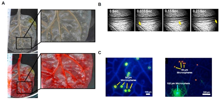
Figure 3.
The 3D Microdevice fabrication Using Spinach Leaf Scaffolds. (A) Optical Image of spinach leaf before and after perfusion; (B) Video frames of microspheres traveling through scaffolding; (C) Fluorescence images of microspheres traveling through scaffolding (adapted from Gershlak et al. [34]).

