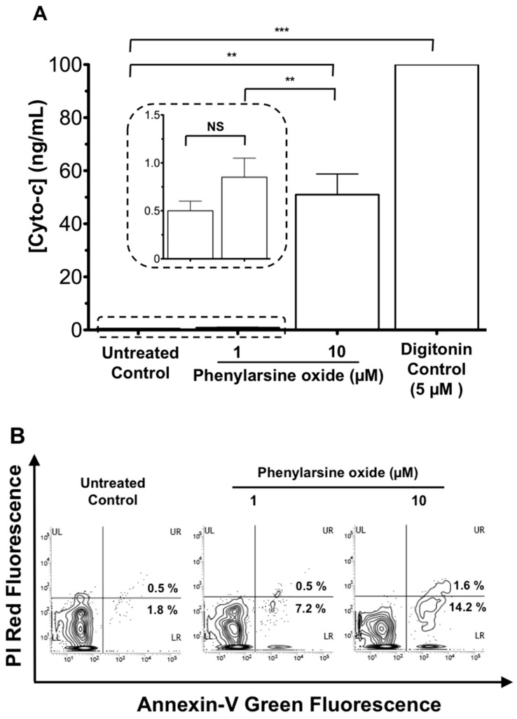
Figure 6.
Cyto-c release during anti-cancer drug treatment using leukemia-sparked samples. HL-60 cells (1 × 10⁵/mL) in serum were cultured with the anti-cancer drug phenylarsine oxide at a concentration as indicated (1, 10 μM) or digitonin (5 μM) as a positive control at 37 °C, 5% CO2 for 1 h. (A) Cell-free sera were then collected by filtering with a 0.2 μm filter unit and subjected to the AuNR-LSPR assay for determining the release of cyto-c. Results are mean ± SEM (n = 3), NS p > 0.05, * p < 0.05, ** p < 0.01, *** p < 0.001. (B) Cells after treatment were labeled with Annexin-V-GFP and PI. After washing, cells were subjected to flow cytometric analysis for green and red fluorescence. The fluorescence properties of 1000 cells were collected for analysis.

