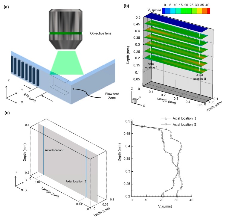
Figure 5.
(a) Schematic illustration of the microfluidic device depicting the observation zone, which is situated at a distance of 270 µm from the terminally located artificial cilia; (b) Calculated ensemble streamwise velocity distribution of the six selected horizontal planes along the depth of the microfluidic device, situated at distances of 200 µm, 250 µm, 300 µm, 350 µm, 400 µm, 450 µm, 500 µm, respectively, from the bottom of the microfluidic device. Two different velocity profiles were evidenced at two axial locations situated at distance of 310 µm (Axial location I) and 710 µm (Axial location II) away from the rearmost artificial cilia; (c) Quantified instantaneous velocity profiles at the two different axial locations (i.e., axial location I and axial location II) of the observation zone along the vertical mid plane of the channel width.

