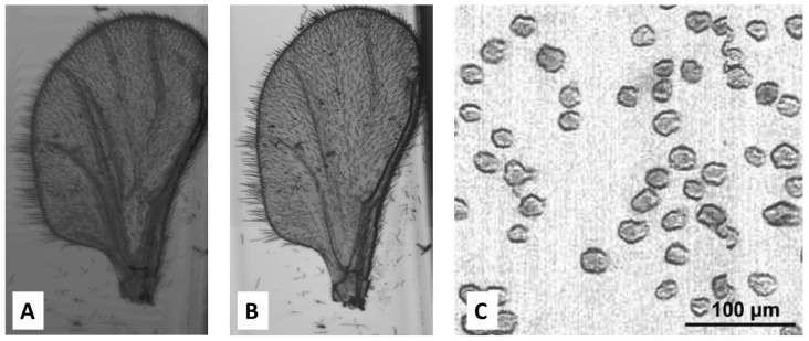
Figure 8.
Comparison of images of a gnat wing obtained from (A) a CD-based system as a microscope and (B) a conventional bright field laboratory microscope. Adapted with permission of Taylor & Francis from [108]. (C) Demonstration of CD player as a diagnostic microscopy tool showing imaging of T2 cells (CD4+ and CD8-). Reproduced in part from [107] with permission of The Royal Society of Chemistry.

