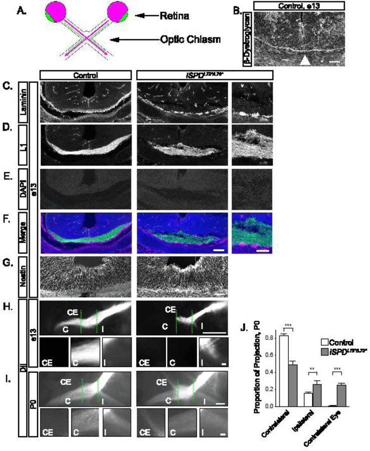
Figure 1: Glycosylated dystroglycan is required for axon guidance at the optic chiasm.

(A) Schematic of RGC axon crossing at the optic chiasm, with contralateral projections in purple, ipsilateral projections in green. (B) At e13, β- Dystroglycan is enriched at the basement membrane in the ventral forebrain (arrowhead). (C-F) Sections at e13 reveal RGC axons (L1, D, green, F) in direct proximity to the basement membrane (laminin, C, purple, F) in controls (left panels). The basement membrane is discontinuous in ISPDL79*/L79* mutants (right panels), and axons are defasciculated and enter the ventral forebrain. DAPI reveals that the cellular architecture around the chiasm is normal (E). (G) The endfeet of Nestin+ midline glia at the optic chiasm appear disorganized in ISPDL79*/L79* mutants compared to controls. (H) At e13, DiI labeled RGC axons form an exclusively contralateral projection at the optic chiasm (dashed green lines) in controls (left). Axons in ISPDL79*/L79* mutants fail to advance though the chiasm and form an abnormal ipsilateral projection (right). (I) At P0, axons in controls make a dominant contralateral and minor ipsilateral projection (left). In ISPDL79*/L79* mutants, axons project contralaterally, ipsilaterally, and to the contralateral eye (right). (J) Quantification of RGC axon trajectories indicate that mutant axons have a reduced contralateral projection (ANOVA, Tukey HSD post hoc test, p<0.01, n=12 control, 6 mutant chiasms). C denotes contralateral projection, I denotes ipsilateral projection, and CE denotes contralateral eye projection. Scale bars: B, 50μm; C-F, 100μm; C-F insets, 50μm; G, 50μm; H, I, 200μm; H insets, 10μm; I insets, 50μm.
