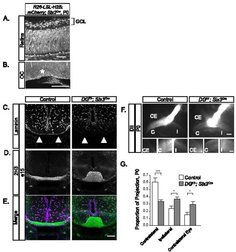
Figure 2: Conditional deletion of dystroglycan results in RGC axon guidance defects at the optic chiasm.

(A-B) Sections from Rosa26-lox-stop- lox-H2B:mCherry;Six3Cre mice show recombination throughout the retina (A) and in cells lining the optic chiasm at P0. (C-E) Sections through the ventral forebrain at e15 reveal a loss of laminin staining at the chiasm (C, E purple) and axons (2H3) grow inappropriately into the ventral forebrain (D, E green) in DGF/-;Six3Cre mice. (F) RGC axons in DGF/-;Six3Cre mutants stall at the optic chiasm and have reduced projections into the contralateral and ipsilateral optic tracts. (G) Quantification of RGC axon trajectories indicate that mutant axons have a decreased contralateral projection and increased ipsilateral and contralateral eye projections compared to controls (ANOVA, Tukey HSD post hoc test, p<0.01, n=4 control, 3 mutant chiasms). C denotes contralateral projection, I denotes ipsilateral projection, and CE denotes contralateral eye projection. Scale bars: A, 50 μm; B, 500μm; C-F, 200μm; F insets, 50μm.
