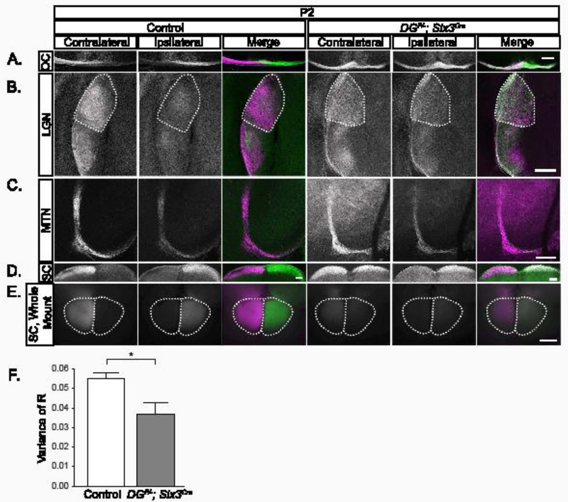
Figure 3: Retinal ganglion cell axons target retinorecipient regions in DGF/-; Six3Cre mice.

(A) CTB labeled axons at the optic chiasm show eye-specific segregation in controls at P2 (left panels) and overlapping projections in DGF/-; Six3Cre chiasms (right panels). (B) Axons from the contralateral retina (purple) innervate the dorsal and lateral LGN, while axons from the ipsilateral retina (green) are restricted to LGN core regions in controls (left) at P2. Axons from the contralateral and ipsilateral retinas overlap across the entirety of the LGN in DGF/- ;Six3Cre mutants. (C) In the MTN, controls and DGF/-;Six3Cre mutants receive innervation from both the contralateral retina (left), and the ipsilateral retina (D, E) In sections (D) and whole mount views of the SC (E), controls (left) are innervated by the contralateral retina. In DGF/-;Six3Cre mice (right), there is innervation from both contralateral and ipsilateral retina across the entire SC. (F) Threshold-independent analysis of LGN projections (B) is measured by the variance of R. Smaller values indicate less segregation and greater axonal overlap, whereas larger values indicate higher segregation and less axonal overlap. The analysis shows a significantly greater axonal overlap in the dLGN in mutants than controls (t-test, p=0.038, n=3 control, 3 mutant). Dotted lines indicate dLGN. Scale bar 200μm.
