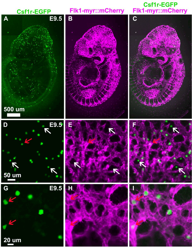
Figure 1.
Csf1r-EGFP+ cells are found in and around the vasculature (Flk1-myr::mCherry+). A., B. and C. Confocal images of an E9.5 Csf1r-EGFP+/tg; Flk1-myr::mCherry+/tg transgenic embryo showing EGFP+ embryonic macrophages dispersed throughout the embryo with greater density anteriorly. D. to I. High magnification images of the anterior dorsal part of the embryonic head at E9.5. EGFP+ embryonic macrophages appear in close association with the vasculature, in both the lumen of the vasculature (red arrow) and extravascular spaces (white arrow), and exhibit a rounded morphology.

