
Figure 5. Thalamic PHb projects to mood-regulating centers.
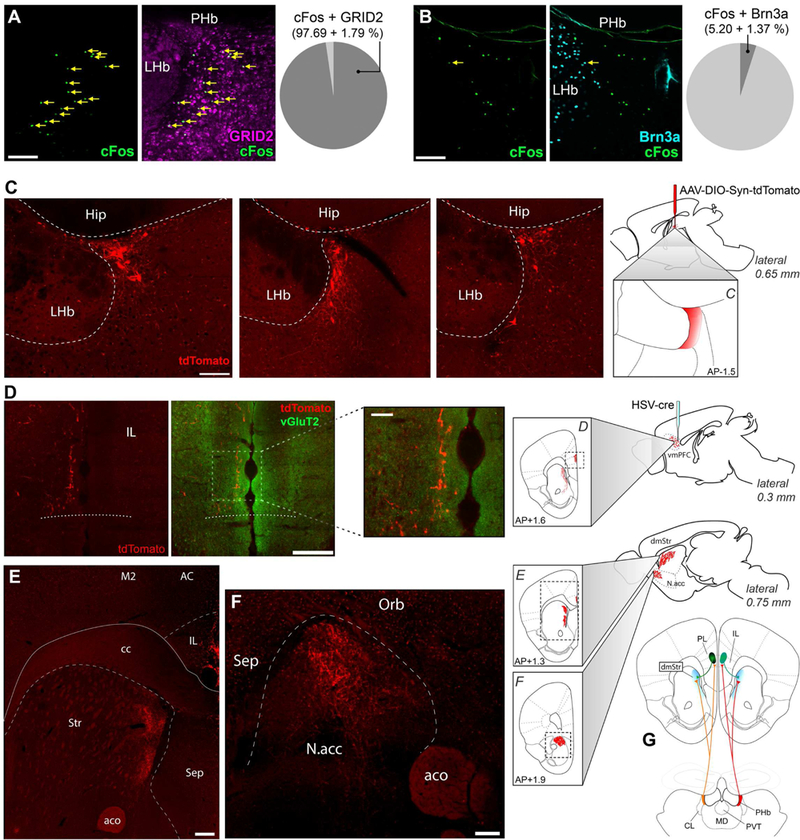
(A-B) Light induced cFos(+) cells in the PHb were colocalized with GRID2 (A) or Bm3a (B) markers. Data are mean±SEM; n=3 mice. (C-F) HSV/cre was injected into the vmPFC, while AAV/DIO-synaptophysin-tdTomato was injected into the PHb. Labeled somas were exclusively found in the PHb (C). PHb neurons have three targets: the vmPFC, including the IL (D), the dorsomedial striatum (E), and the NAc (F). n=12 mice. (G) These experiments revealed a thalamocortical loop, represented here diagrammatically. cc: corpus callosum; M2: secondary motor cortex; AC: anterior cingulate cortex; Str: striatum; aco: anterior commissure; Sep: septal nuclei; Orb: orbitofrontal cortex, Hip: hippocampus. Scale bars: (D insert) 50μm; (A, B, C) 100μm; (D, E, F) 200μm. See also Figure S5.