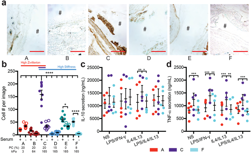
Figure 4. Macrophages adhere better to implants with a more severe foreign body response.
a) Representative images for macrophages stained around the implant, where the # denotes the location of the implant (scale 250 μm). b) Cell adhesion to hydrogels either treated for 30 minutes with serum proteins or not and then seeded with macrophages and imaged 24 hours later (N=2, n=4). Stats displayed for plus and minus serum on hydrogels and hydrogel C plus serum compared to all other hydrogels. Secretion of c) IL-10 and d) TNF-alpha from macrophages seeded on hydrogels with different stimulation factors. Stimulation of each condition was as follows: NS: no stimulation, LPS/IFN-γ: 1ng/mL LPS and IFN-γ, IL4/IL13: 20ng/mL IL4 and IL13, LPS/IL4/IL13: 0.5ng/mL LPS and 20ng/mL IL4 and IL13. The percentage of PC content and the Young’s modulus for each hydrogel is labeled below. Significance is determined using an ANOVA with Tukey’s post-test where p=0.05 is significant. Error bars are the SD (N=2, n=5).

