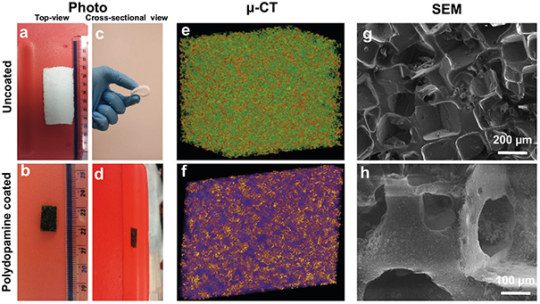
Fig. 2.

Photographs of the top-view (a,b) and cross-sectional view (c,d) of uncoated- (a,c) and polydopamine coated-(b,d) PDMS bioscaffolds showing the macrostructure and the color change from white to brown with polydopamine coating. The reconstructed μ-CT (e,f) and SEM images (g,h) of uncoated-(a,c,e,g) and polydopamine coated- (b,d,f,h) PDMS bioscaffolds
