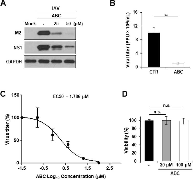
Fig. 3. SphK2 inhibitor suppresses IAV replication in vitro.

(A) A549 cells were infected with IAV at an MOI of 1. Cells were left untreated or treated with ABC294640 (ABC) at different concentrations as indicated. At 24 hpi, the levels of M2, NS1, and GAPDH were analyzed by Western blotting. The experiment was independently repeated twice. (B) A549 cells were treated with solvent control (CTR) or ABC (50 µM). At 1 hour post-treatment, cells were infected with IAV at an MOI of 0.5. The titer of infectious IAV in the supernatants of the culture was assessed by plaque assay on MDCK cells at 24 hpi (n = 3/group; **, p ≤ 0.01). (C) A549 cells were infected with IAV at an MOI of 0.001 for 1 hour. Infected cells were then treated with ABC at 6 different concentrations (0.032, 0.16, 0.8, 4, 20, or 100 µM) or left untreated. Virus titer in the supernatant was measured at 48 hpi by plaque assay on MDCK cells. No inhibition was seen when cells were treated with ABC at 0.032 µM (virus titer = 2.3 × 10⁶ PFU/mL). The EC50 was determined with GraphPad Prism 5 software. The result represents the average of 3 replicative experiments. (D) A549 cells were treated with solvent (−) or ABC (20 µM or 100 µM) for 48 hours. Cellular viability was monitored by using a trypan blue exclusion assay. The total number of live cells in untreated group was set as 100%, and the relevant number of live cells in the ABC-treated groups are shown in percentages. The data represent means ± SD (n=3). n.s. = not significant.
