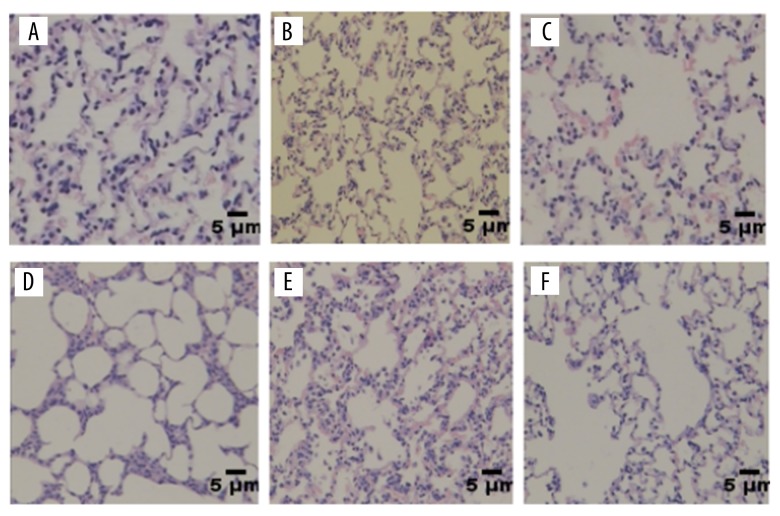
Figure 1.
Analysis of lung tissue hematoxylin and eosin staining. There was no significant change in lung alveolar area and alveolar septum in the normal control group (A, B). The alveolar area in lung tissue was slightly increased and the alveolar septum thickened and ruptured in the IH 12-week group (C). The lung tissue damage was more severe in the IH 16-week group (D). The degree of destruction of the lungs in the 12-week HI group was lower than in the 12-week IH group (E). The degree of lung destruction in the HI 16-week group was lower than in the IH 16-week group (F).

