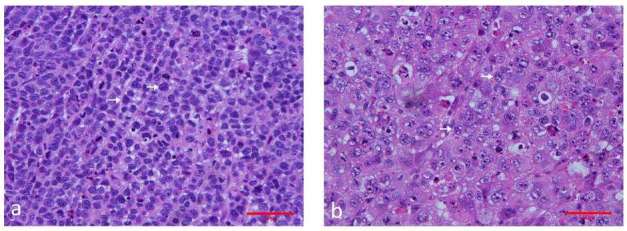
Figure 5. .

Representative images of hematoxylin and eosin–stained sections of the control and treated xenograft breast tumor in nude mice. (a) xenograft tumor of the control group. The tumor cells are tightly packed and most have large darkly stained nuclei. Original magnification, ×400. (b) xenograft tumors treated with cisplatin (2 mg kg–1) + paclitaxel (10 mg kg–1) for 5 days. The tumor tissue shows marked regression, steatosis, apoptosis and cell debris. Original magnification, ×400.
