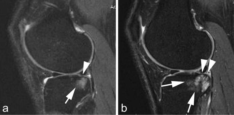
Figure 3.

Structural predictors of progression. (a) Baseline sagittal intermediate-weighted fat-suppressed MRI shows minimal superficial focal cartilage defect at posterior lateral tibia (arrow head). In addition there is an associated large diffuse subchondral bone marrow lesion (arrow). (b) Follow-up MRI 2 years later shows progression of cartilage damage with now diffuse cartilage loss at the posterior tibia (arrow heads) and further progression of subchondral bone marrow lesion (arrows). Bone marrow lesions are strong predictors of subsequent structural progression including cartilage loss.
