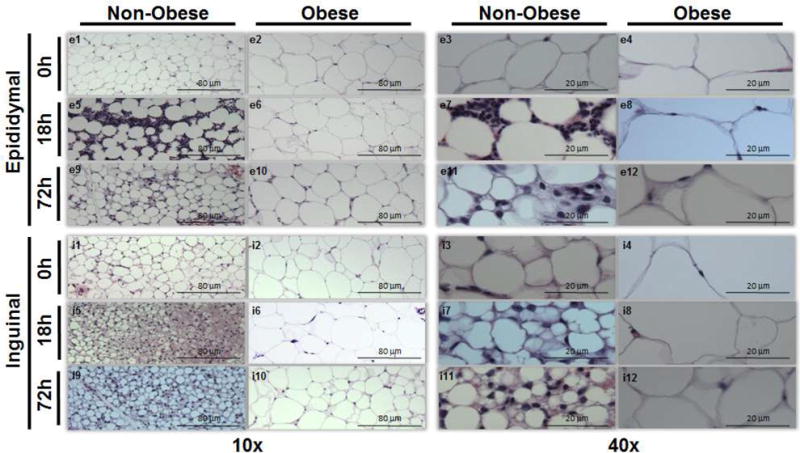
Figure 2. WAT H&E staining from epididymal and inguinal WAT.

Epididymal WAT in non-obese mice at 0h (e1,3), 18h (e5,7), and 72h (e9,11) and in obese mice at 0h (e2,4), 18h (e6,8), and 72h (e10,12) after sepsis at 10× and 40× magnification respectively. Inguinal WAT in non-obese mice at 0h (i1,3), 18h (i5,7), and 72h (i9,11) and in obese mice at 0h (i2,4), 18h (i6,8), and 72h (i10,12) after sepsis at 10× and 40× magnification respectively. Representative sections are illustrated. A similar pattern was seen in n=2-4 different samples in each experimental group. All mice were 12 weeks of age at the time of harvest.
