
Figure 4. Transmission electron microscopy of epididymal WAT in a non-obese mouse 18 hours after CLP.
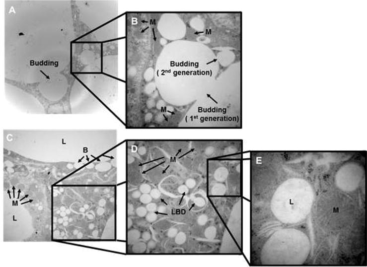
(A) and (B) demonstrate the budding process in which lipid droplets breakdown into 1st and 2nd generation lipid droplets. (C), (D) and (E) show the close proximity between the newly formed small lipid droplets and mitochondria at 1,500×, 3,000×, 8,000× respectively. Mice were 12 weeks of age at the time of harvest. Representative sections are illustrated. A similar pattern was seen in n=2-4 different samples. L=lipid droplets, B=budding of the lipid droplets, LBD=lipid breakdown, M=mitochondria.