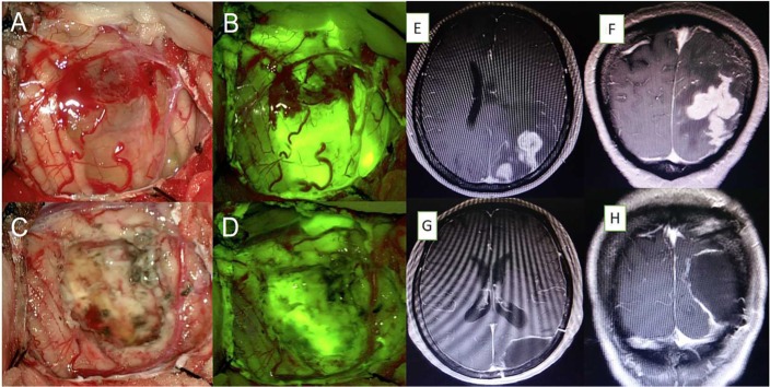
Figure 1.
(A) Microsurgical photograph of the right parietal lobe under white light and (B) the yellow 560 filter from initial stages of tumor resection. (C, D) Residual tissue located in the deep area of the resection cavity was identified under fluorescence compared to white light. (E–H) Axial and sagittal magnetic resonance imaging showing complete removal of the tumor.

