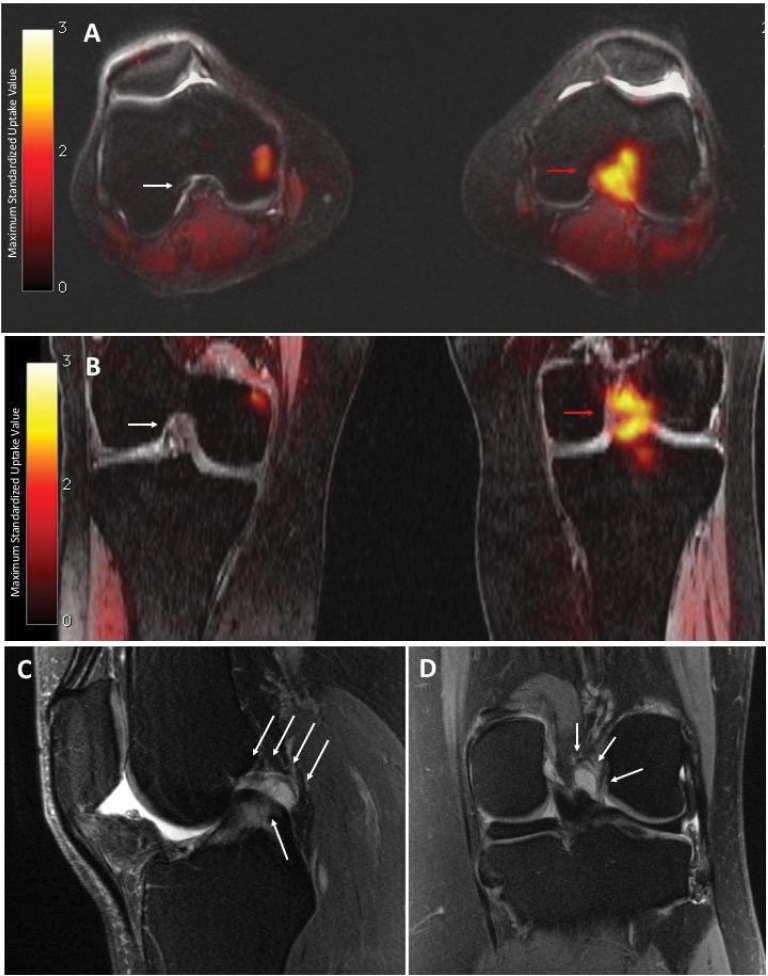
Figure 1.
PET/MRI and MRI of the left knee
Notes: Axial (A) and coronal (B) PET/MRI show high uptake of 18F-FTC-146 in the intercondylar notch (red arrows: maximum standardized uptake value = 2.04). By comparison, the intercondylar notch of the right knee is normal (white arrow: maximum standardized uptake value = 0.17). MRI shows small joint effusion, with no synovitis, and an amorphous, mass-like in the intercondylar notch, initially presumed to be a ganglion cyst or localized pigmented villonodular synovitis/fibrous lesion. Sagittal (C) and coronal (D) MRI (T2-weighted with fat saturation) of the left knee, acquired approximately 3 years before PET/MRI study, show abnormal high-signal amorphous, mass-like but equivocal lesion in the intercondylar notch (white arrows). This had been overlooked or regarded as clinically insignificant.

