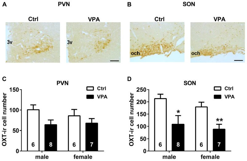
FIGURE 3.

Neonatal VPA rats had fewer OXT-ir cells in the hypothalamus than controls. Representative images (bar = 100 μm) of OXT staining in the PVN (A) and SON (B) of both control and VPA rats. In the PVN (C), the number of OXT-ir cells did not differ between groups. But in the SON (D), they were less in the VPA rats than controls. Data are expressed as mean ± SEM (control: male n = 6, female n = 6; VPA: male n = 8, female n = 7), ∗P < 0.05, ∗∗P < 0.01. Ctrl, control; PVN, paraventricular nucleus; SON, supraoptic nucleus; 3v, third ventricle; and och, optic chiasm.
