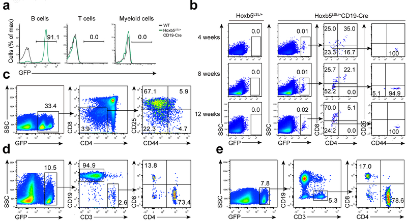
Figure 3. Conversion of B to T lymphocytes using a CD19-Hoxb5 transgenic model.
(a) GFP reported B-lineage specific expression of ectopic Hoxb5 in the PB of Hoxb5LSL/+ CD19-Cre compound (CD19-Hoxb5) mice. CD19+ B cells, CD3+ T cells, and Mac1+ Myeloid cells were analyzed by flow cytometry. Numbers above bracketed lines indicate percent GFP+ cells, (b) Flow cytometry analysis of thymic Ter119−Mac1−CD19−GFP+ lymphocytes of 4, 8, and 12 weeks old CD19-Hoxb5 mice and littermate control mice under homeostasis. (c-e) Flow cytometry analysis of iDN cells in the thymus gated from Ter119−Mac1−CD19− population (c), iT cells in spleen (d), and LN (e) gated from Ter119−Mac1− population of a representative recipient transplanted with three million CD19-Hoxb5 pro-pre-B cells four weeks after transplantation. Data are representative of two independent experiments (a, b) or three independent experiments (c, d and e).

