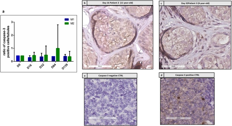
FIGURE 5.
Active caspase 3 immunostaining. (a) Shows the evolution of intratubular cells stained for active caspase 3, which did not show any significant change along the culture. (b,c) Show the staining of caspase 3 in patient 2 at day 16 (b) and for patient 3 at day 32 (c), where no stained cells were detected in the adluminal compartment of corresponding STs presented on HE sections in Figures 8c,d. Some positive cells are found at the basement membrane. (d,e) Show negative and positive controls for caspase 3 performed on tonsil tissue.

