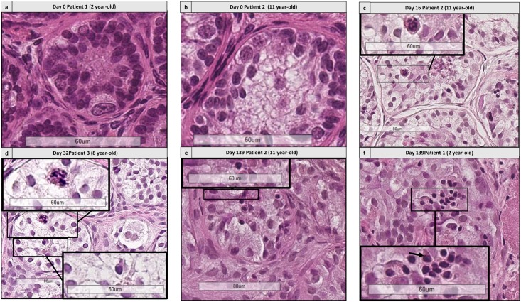
FIGURE 8.
Histological assessment of differentiating germ cells. (a,b) Shows the presence of spermatogonia from patient 1 aged 2 years old and patient 2 aged 11 years old. (c–f) Shows the presence of differentiating germ cells at early culture time points (D16 and 32) and late time points (D139) for patient 2 (c,e) aged 11 yearss old, for patient 3 aged 8 years old (d) and patient 1 aged 2 years old, culture medium 2 (f). Cutouts show the differentiating germ cells at a higher magnification, like a spermatocyte with a characteristic granulo-filamentous chromatin (c,d), and round spermatids with the small condensed and eccentric nucleus, adluminal localization and pear shape (d–f).

