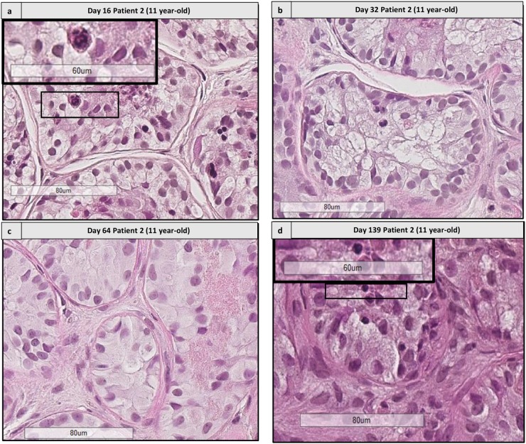
FIGURE 9.
Histological assessment of sequential images from the same patient. This figure shows the histological images from patient 2 (aged 11 years old) from days 16 to 139. (a,d) Shows the presence of differentiating cells at days 16 and 139, respectively. Cutouts show the differentiating germ cells at a higher magnification, like a spermatocyte with a characteristic granulo-filamentous chromatin (a), and round spermatids with the small condensed and eccentric nucleus, adluminal localization and pear shape (d). In the timepoints 32 and 64 (b,c) we did not find any differentiating cells, as different fragments were harvested for the different timepoints.

