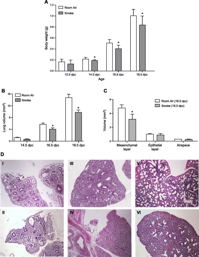
Figure 1.

A) Whole body weight measurements of room air– and smoke-exposed fetal lungs. Significant decrease in embryonic body weight due to maternal smoke is detectable beginning at 14.5 dpc. B) Comparative lung volume measurements of room air– and smoke-exposed fetal lungs. Cavalieri estimations reveal significant decrease in fetal lung volume due to maternal smoke beginning at 16.5 and 18.5 dpc. C) Mesenchymal, epithelial, and airspace volumes of room air– and smoke-exposed 16.5 dpc fetal lungs. Smoke-induced reduction in lung volume at 16.5 dpc is attributable to decrease in lung mesenchyme (room air: 4.78 ± 0.47 mm3; smoke: 3.17 ± 0.73 mm3; *P = 0.032). Asterisks denote significant difference in means at P ≤ 0.05 by Student’s t test. D) Representative images of lung tissue slices obtained from room air– and smoke-exposed mice. Serial sections (5 μm) of paraffin-embedded lungs were used to obtain measurements of mesenchymal, epithelial, airspace, and total lung volumes. Lung slices of room air–exposed fetal mice at 14.5 (I), 16.5 (III), and 18.5 (V) dpc are displayed, along with comparative lung slices of in utero smoke-exposed mice at 14.5 (II), 16.5 (IV), and 18.5 (VI) dpc. Original magnification, ×20.
