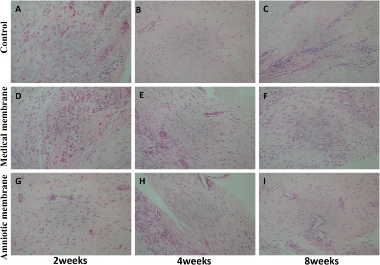
Fig 4. Histological observation of the tendon sheath defection in each group at the 2nd, 4th, and 8th week after operation (hematoxylin–eosin staining, x 400).
At the 2nd week, each group was filled with hyperemia, edema, and inflammatory cell infiltration, but the amniotic membrane group (G) was the lightest. At the 4th week, inflammatory cells were significantly reduced than before. In the medical membrane group (E) and the amniotic membrane group (H), fibroblast cells were layered, and the amniotic membrane group was dense. In the control group (B), numerous fibroblasts were disorderly distributed. At the 8th week, the fibroblasts in the medical membrane group (F) and the amniotic membrane group (I) were arranged neatly, and the structure was dense. In the control group (C), the structure of tendon sheath was loose, and the distribution of fibroblasts was disorganized.

