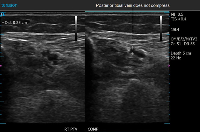
Fig. 3.

This 49-year-old woman returned in follow-up 6 days after surgery. Her ultrasound scan demonstrated a thrombus in a right posterior tibial vein. There was no evidence of popliteal extension. This image shows noncompression of one of the posterior tibial veins, indicating the presence of an intraluminal mass. The patient’s color flow and waveform images are shown in Figures 4, 5.
