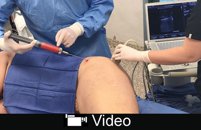
Video Graphic 1.

See video, Supplemental Digital Content 1, which displays an intraoperative video of a 44-year-old woman undergoing fat injection of the left buttock. The patient is positioned on her right side. Fat harvesting has already been completed. The patient had liposuction and an abdominoplasty. The monitor shows the cannula within the subcutaneous fat layer. Fat can be seen exiting the cannula (red circle), well above the gluteus maximus muscle fascia. The 2-second ultrasound imaging segment is shown at normal speed, but repeated ×7 to allow the reader enough time to view the fat escaping from the end of the cannula, http://links.lww.com/PRSGO/A838.
