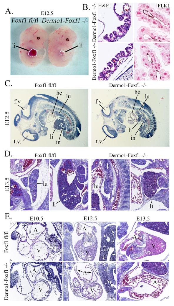
Figure 2. Developmental abnormalities in Dermo1-Foxf1−/− embryos at E12.5-E13.5.
A, Representative photographs of Dermo1-Foxf1−/− and control Foxf1fl/fl embryos show no differences in body sizes at E12.5. B, H&E and immunostaining for FLK1 show no vascular abnormalities in yolk sacs of E13.5 Dermo1-Foxf1−/− embryos. FLK1-stained slides (dark brown) were counterstained with nuclear fast red (red nuclei). C-E, H&E staining of E10.5-E13.5 embryos show lung and liver hypoplasia (C-D) and heart defects (E) after deletion of Foxf1. Abbreviations: lu, lung; li, liver; in, intestine; he, heart; A, heart atrium; V, heart ventricle; t.v., third brain ventricle; f.v., fourth brain ventricle. Magnification: B (left panels), x200; B (right panels), x400; C, x16; D and E (middle and right panels), x50; E (left panels), x100.

