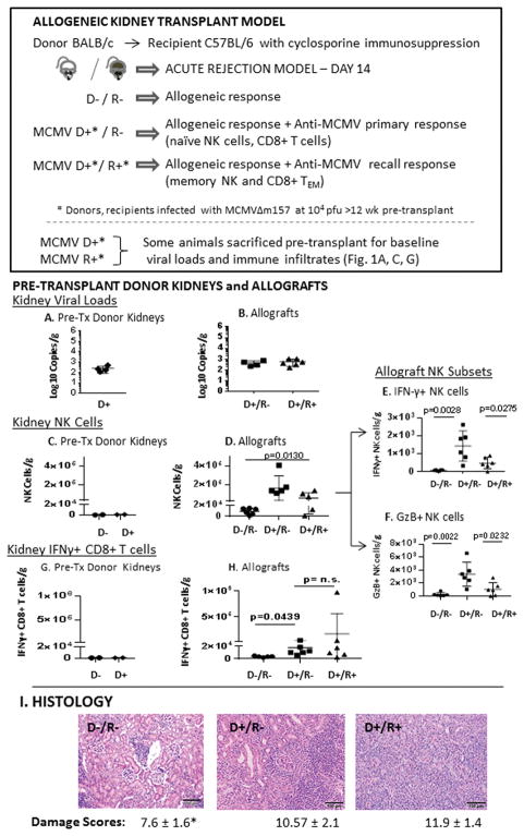
FIGURE 1. NK and CD8+ T cell primary and memory responses in MCMV infected allografts.
Donor BALB/c and recipient C57BL/6 (B6) mice were infected with MCMVΔm157 strain at 104 pfu by intraperitoneal injection at least 12 weeks prior to transplantation to establish NK and CD8+ T cell antiviral memory. MCMV D−/R−, D+/R−, and D+/R+ allogeneic kidney transplants were performed with cyclosporine immunosuppression, and allograft-infiltrating leukocytes were analyzed by flow cytometry on post-transplant day 14. MCMV infected BALB/c and B6 mice were sacrificed > 12 weeks post-infection to establish baseline pre-transplant organ viral loads and leukocyte infiltrates.
(A, B) DNA was extracted from (A) pre-transplant MCMV+ BALB/c kidneys or (B) post-transplant D+/R− and D+/R+ allografts, and MCMV viral loads were determined by quantitative DNA PCR.
(C–F) Kidneys from (C) pre-transplant uninfected (D−) or MCMV infected (D+) BALB/c kidneys, or (D) allografts from D−/R−, D+/R− and D+/R+ transplants were processed for total live cells gated on CD45+/MHCII-/CD3-/NKp46+ NK cells, and the number of NK cells expressing (E) interferon-γ (IFN-γ+ NK cells) or (F) granzyme B (GzB+ NK cells) were compared between groups.
(G, H) Total live cells from (G) pre-transplant uninfected (D−) or MCMV infected (D+) BALB/c kidneys, and (H) D−/R−, D+/R− and D+/R+ allografts were gated on CD45+/MHCII-/CD3+/CD8+ T cells expressing IFN-γ and compared between groups.
(I) Allograft tissues from D−/R−, D+/R−, and D+/R+ transplants were fixed in formalin, paraffin embedded, and stained with hematoxylin and eosin. Allograft damage was graded using an 8-criteria scale (maximum score 24), and scores were compared between groups (n=6–8/group; * p<0.05). Images were collected under identical conditions at 20x magnification. Scale bar=100 μm.

