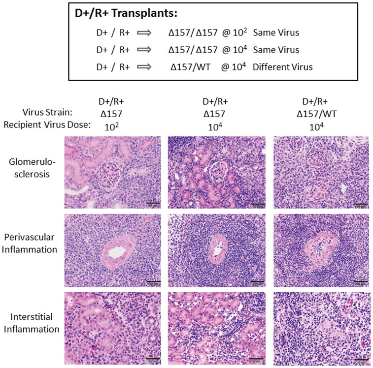
FIGURE 9. Allograft histopathology from MCMV immune recipients.
D+/R+ transplants were performed using MCMVΔ157 same-strain infections for recipients with low dose (102) or high dose (104) infections, and compared to D+/R+ MCMVΔ157/MCMV WT different strain infections (104), as described in Figures 3 and 7. At day 14 post-transplant, allografts were stained with hematoxylin and eosin and allograft damage scored according to 8 histologic criteria (Table 1). Representative images for glomerulosclerosis, arteritis, and interstitial inflammation were collected under identical conditions at 40x. Scale bar=50 μm.

