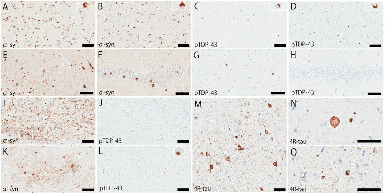
Figure 4.
Immunohistochemistry for α-synuclein (A, B, E, F, I, and K) and phospho-TDP-43 (C, D, G, H, J, and L). The mammillothalamic tract (A and C), thalamic fasciculus (B and D), hippocampal CA1 (E and G), dentate gyrus (F and H), transverse fibers in pontine base (I and J), and inferior olivary nucleus (K and L). Immunohistochemistry for 4-repeat tau shows representative pathologic features of argyrophilic grain disease, including argyrophilic grains (M), pretangles (M), and balloon neurons (N) in the amygdala, and coiled bodies (O) in the white matter in the parahippocampal gyrus. Bars: 100 μm in A–L, and 50 μm in M–O.

