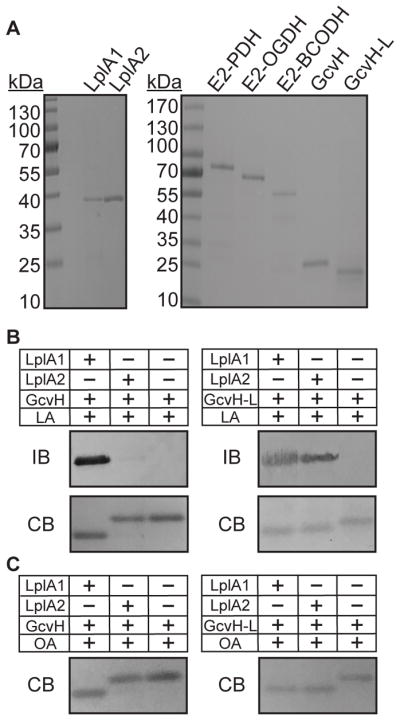
Figure 3. Targeting of GcvH and GcvH-L by LplA1 and LplA2.

(A) GelCode Blue stained SDS-PAGE gels containing 2 μg of the salvage enzymes, LplA1 and LplA2, and lipoyl domain containing subunits E2-PDH, E2-OGDH, E2-BCODH, GcvH, and GcvH-L. (B–C) LplA1 and LplA2 attachment of (B) lipoic acid (2.4 mM) and (C) octanoic acid (2.4 mM) to GcvH and GcvH-L. Lipoylation was assessed by conducting an immunoblot (IB) with rabbit α-lipoic acid antibody. Parallel 12% SDS PAGE gels were stained with GelCode Blue (CB). Octanoylation was visualized as a shift in apparent molecular weight after resolving proteins on a 12% SDS-PAGE gel and staining with GelCode Blue (CB).
