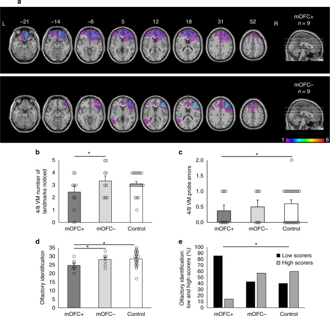
Fig. 5.
Patients with medial orbitofrontal cortex (mOFC) damage show reduced spatial memory. a Axial slices and midsagittal view of the ICBM52 MNI brain depicting the extent of the lesion overlap for patients whose frontal lobe lesions encompass the mOFC (mOFC + ; top) and patients whose frontal lobe lesions do not encompass the mOFC (mOFC-; bottom). The numbers above the axial slices indicate their z coordinates in MNI space. The colors indicate the degree to which lesions overlap within each group, as indicated by the color scale, which ranges from 1 to 8 patients. b In the 4/8 VM, the mOFC + group noticed fewer landmarks in the environment compared to the mOFC- group (mean difference = −1.17, Bootstrap BCa 95% CI [−2.20, −0.27]). c The mOFC + group also made fewer errors on the probe trial compared to the control group (mean difference = −0.24, Bootstrap BCa 95% CI [−0.55, −0.02]). d The mOFC + group exhibited worse olfactory identification than both the mOFC- group (mean difference = −4.99, Bootstrap BCa 95% CI [−8.54, −1.09]) and the control group (mean difference = −3.70, Bootstrap BCa 95% CI [−6.02, −1.12]). e We categorized participants as low (black bars) or high (gray bars) scorers on the olfactory indentification task according to the median across all groups (median = 27.5). The mOFC + group included more low scorers than the control group, p < 0.05 (Fisher’s exact test). These results indicate that mOFC damage affects both spatial memory and olfaction, as it is is associated with a lesser reliance on landmarks and worse olfactory identification. Error bars indicate the standard error of the mean. *indicates significance based on bootstrapped bias-corrected 95% confidence intervals. L Left, R Right

