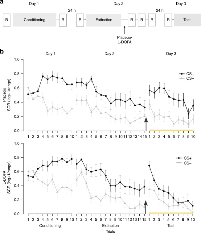
Fig. 1.
Experimental design and skin conductance responses. a Participants underwent a 3-day fMRI study with fear conditioning on day 1, extinction and subsequent placebo or l-DOPA administration on day 2 and test on day 3. During fear conditioning one of two geometric symbols (CS+) was reinforced with a painful electrical stimulation, while the other symbol (CS−) was never reinforced. Postextinction placebo or l-DOPA administration was randomized and double-blinded (placebo: n = 20, l-DOPA n = 20, all male, for group characteristics, see Supplementary Table 1). Resting-state fMRI scans (R) were acquired before and after fear conditioning, before and 10, 45, and 90 min after extinction, and before test. b During all experimental phases, we assessed conditioned responses (CRs) as skin conductance responses (SCRs) to CS+ and CS−. Upper panel depicts mean SCR to CS+ and CS− for placebo-, lower panel for l-DOPA-treated participants. The groups differed significantly on mean SCRs across the test phase on day 3 (marked by yellow line) due to significantly smaller mean SCRs to the CS+ in l-DOPA compared to placebo-treated participants. Note, that the group difference stemmed from significantly smaller CS+ evoked SCRs averaged across the whole test phase, but the speed of re-extinction did not differ significantly between drug groups (control analysis with stimulus (CS+, CS−) and trial (1–10) as within-, and group (placebo, l-DOPA) as between-subject factor: stimulus × group, F1,33 = 6.58, P = 0.02, partial η2 = 0.17; stimulus x trial x group, F9,297 = 1.32, P = 0.23; n = 35). Data are presented as mean ± standard error of the mean (s.e.m)

