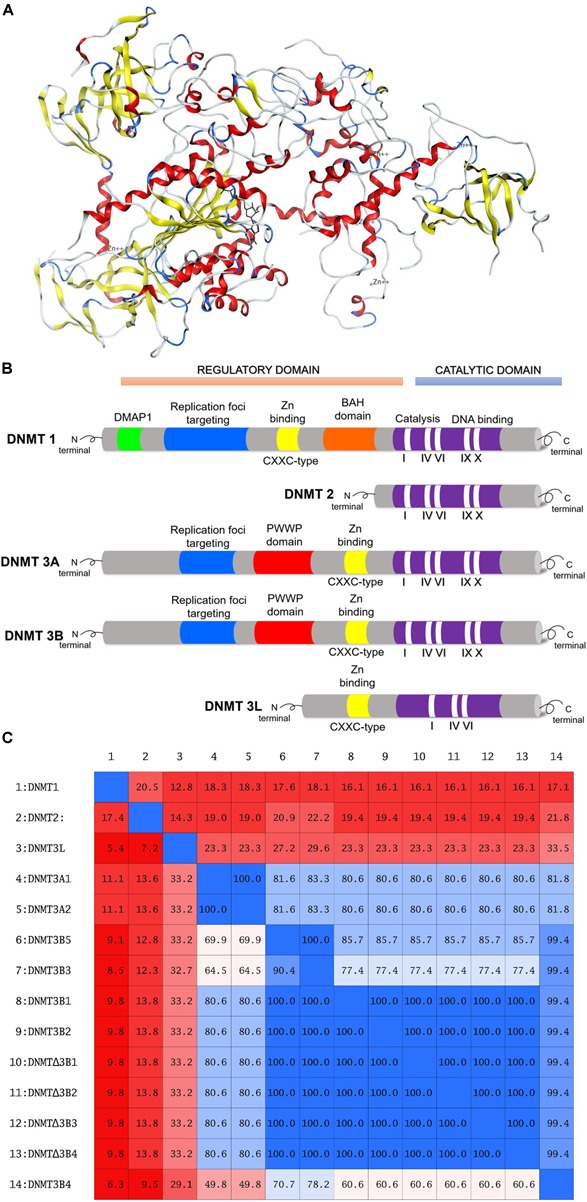
FIGURE 1.

(A) Three-dimensional model of DNMT1, amino acid residues 351–1600. Figure rendered from the Protein Data Bank PDB ID: 4WXX. (B) Schematic diagram of the structure of human DNMT1, DNMT2, DNMT3A, DNMT3B, and DNMT3L. (C) Identity matrix of the catalytic site of 14 DNMTs isoforms. Note that there is a significant difference in the sequence of DNMT1, DNMT2, and DNMT3L.
