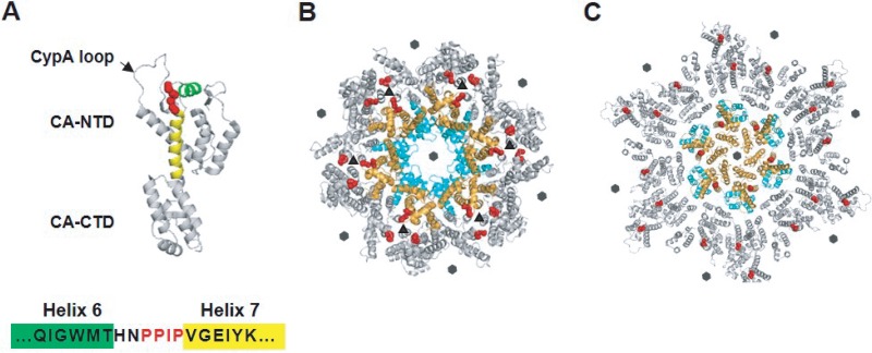
FIG 1.
Location of the PPIP(122–125) motif in the HIV-1 CA monomer, immature Gag lattice, and mature capsid. (A) HIV-1 CA is composed of N-terminal and C-terminal domains (CA-NTD and CA-CTD, respectively) connected by a flexible linker. The loop that binds the cellular protein CypA is indicated (CypA loop; arrow). The PPIP motif (red) is located between helices 6 (green) and 7 (yellow) in the CA-NTD (PDB ID: 5MCX [27]). (B) The CA domain is arranged in a hexameric fashion in the immature Gag lattice (in the central hexamer CA-NTDs in orange and CA-CTDs in cyan; neighboring hexamers in gray) (PDB ID: 4USN [21]). The PPIP motifs (red) of neighboring hexamers meet in the 3-fold interhexamer interface (triangles). Sixfold symmetry axes are indicated by hexagons. (C) A mature hexameric lattice in the intact virion (in the central hexamer, CA-NTDs in orange, CA-CTDs in cyan, neighboring hexamers in gray). Conformational shifts during maturation move the PPIP motif (red) away from the interhexamer interface in the immature Gag lattice to a more central position in the mature CA lattice (PDB ID: 5MCX [27]). Sixfold symmetry axes are indicated by hexagons.

