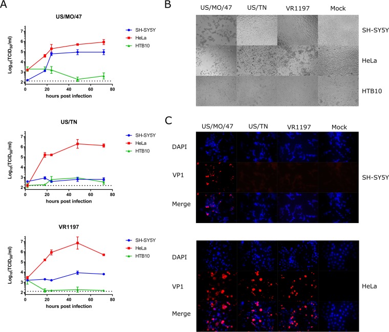
FIG 1.
Differential infection and replication of EV-D68 strains in SH-SY5Y. (A) SH-SY5Y, HTB10, and HeLa cells were grown to 90% confluence in 96-well plates before infection with EV-D68 US/MO/47, US/TN, and VR1197 at an MOI of 0.1. Infection medium was removed 2 h postinfection (hpi) to reduce background. Cell culture lysates were collected at various time points after infection, and viral titers were measured using endpoint dilutions for growth in HeLa cells. The dotted black line indicates the limit of detection. Error bars represent standard error of the mean (SEM) from three biological replicates. (B) SH-SY5Y, HTB10, and HeLa cells were infected with EV-D68 US/MO/47, US/TN, and VR1197 at an MOI of 0.1 as described above. Cells were visualized at 72 hpi with bright-field microscopy at 400×. (C) HeLa and SH-SY5Y cells were infected with the indicated EV-D68 strains at an MOI of 1.0. Cells were fixed at 18 hpi and stained with polyclonal antiserum against EV-D68 VP1 (red) and counterstained with DAPI (blue) for detection of nuclei.

