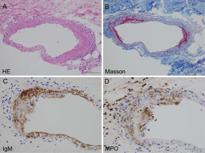
Figure 1.
Light microscopy of a skin purpura biopsy specimen. Light microscopy of a skin purpura biopsy specimen revealed fibrinoid necrosis with neutrophils infiltrating the cutaneous small arteries. (A) Hematoxylin and Eosin staining (×400). (B) Masson staining (×400). Immunostaining revealed (C) IgM and (D) MPO on the vascular walls.

