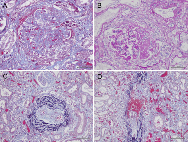
Figure 2.
Light microscopy of a kidney specimen. At autopsy, a pathological examination of the kidney revealed global sclerosis in 50% of the total glomeruli with (A) cellular or fibrocellular crescents and (B) MPGN-like lesions in some parts of the glomeruli. (C, D) The interlobular arteries and arterioles were affected by fibrinoid necrosis with neutrophilic infiltration.

