Figure 5.
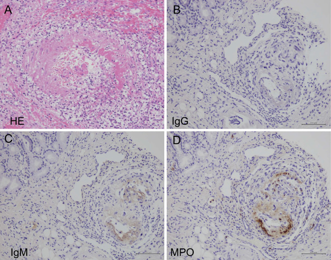
Light microscopy of the gastric mucosa. Light microscopy of gastric and duodenal ulcers revealed fibrinoid necrosis, with neutrophils infiltrating the submucosal small arteries in (A) Hematoxylin and Eosin staining (×400), with positive staining of (C) IgM and (D) MPO on the vascular walls.

