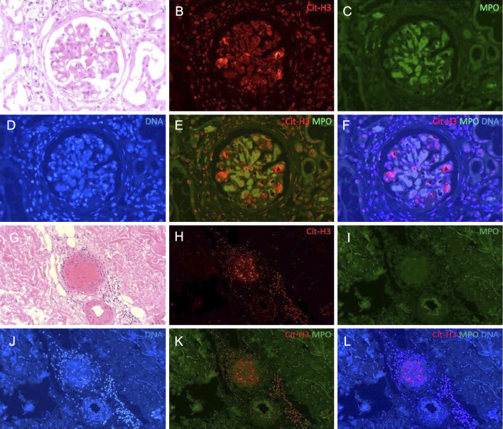
Figure 6.
The immunofluorescence findings of NETs in the kidney and skin purpura. The other lesions of glomerulitis (A-F) and cutaneous small arteries of the skin purpura (G-L). (A, G) Hematoxylin and Eosin staining; (B, H) Cit-H3 staining with anti-Cit-H3 and Alexa Fluor 594-conjugated goat anti-rabbit IgG H&L (red); (C, I) MPO staining with FITC-conjugated anti-MPO (green); (D, J) DNA staining with DAPI (blue). Cit-H3 and extracellular DNA were co-localized with MPO in the glomeruli (F), but not in the cutaneous arteries (L). The small arteries of the gastric and duodenal ulcers were negative for Cit-H3.

