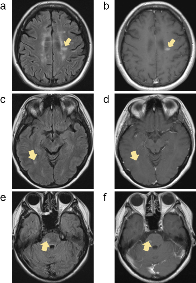
Figure 1.

Magnetic resonance imaging findings of group I. In patient 1, a new gadolinium (Gd) -enhanced lesion was found on magnetic resonance imaging (MRI) at one month after the initiation of dimethyl fumarate (DMF) (a, b). Patient 2 experienced clinical relapse with new Gd-enhanced lesions 98 days after fingolimod cessation (c, d). Patient 3 experienced clinical relapse with new Gd-enhanced lesions 97 days after fingolimod cessation (e, f). All arrows show new lesions with Gd enhancement after fingolimod cessation (a, c, e; FLAIR, b, d, f; Gd-enhanced T1-weighted image).
