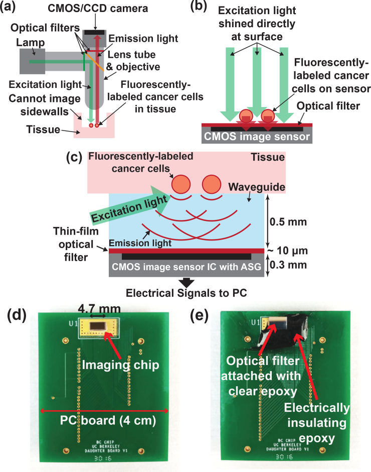
Fig. 1.
(a) Conventional fluorescence microscope. (b) In vitro contact imaging. (c) In vivo contact imaging using an image sensor with ASGs to recover the lost resolution due to light divergence. (d) Image sensor with ASGs bonded onto a PCB. (e) Optical filter epoxied using clear optical epoxy on top of image sensor with ASGs. Dark epoxy is used for electrical isolation.

