Fig. 1.
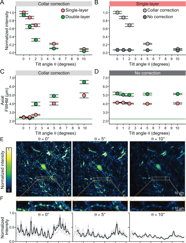
Two-photon fluorescence intensity and FWHM profiles of fluorescent microbeads under different orientation angles, cover glass layers and collar-corrections. (A) Increasing the tilt angle between the optical axis of the two-photon microscope and the axis perpendicular to the imaging window significantly decreases light intensity measured from individual microbeads with a collar corrected objective. Even at small tilt angles of θ = 1° a significant loss of intensity is detected. This effect is amplified when the thickness of the cover glass is doubled. (B) The lack of collar correction drastically decreases the measured bead intensity. (C) With a collar correction, the axial FWHM is fully corrected at θ = 0° tilt angle close to the theoretical limit (green solid line) for single and double cover glass preparations. Increasing the tilt angle significantly degrades the axial resolution. (D) Without collar correction the axial resolution is deceased. The double glass configuration further degrades the axial resolution. The resolution of the uncorrected condition is comparable to a θ = 5° tilt in the corrected condition. (E) Z projection (maximal intensity) of 7 slices (14 µm) at the soma level of a layer 2/3 neuron imaged in the frontal cortex at a 5x magnification factor. The same neuron was imaged at different tilt angles (θ = 0°, 5°, and 10°). (F) Top: zoom in of a neuronal process with boutons acquired at 15× magnification factor. The maximal intensity Z projection (15 slices, 15 µm) was used to obtain the intensity profile of the region of interest on 3 different acquisitions at each tilt angle. Bottom: intensity profiles were aligned to the peaks (thin traces) and averaged (thick traces) for comparison across conditions. Scalebar: 10µm

