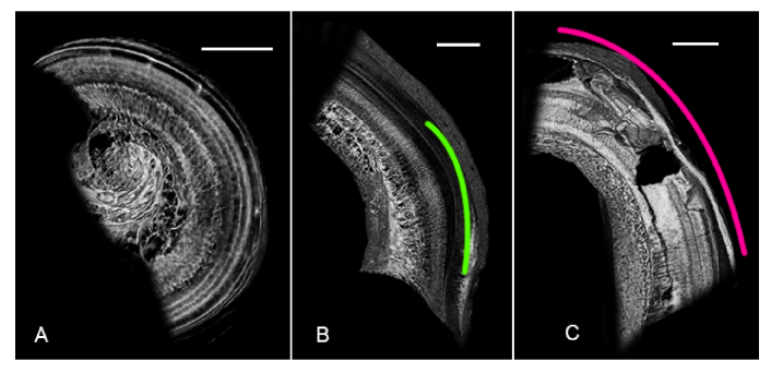
Fig. 6.
Virtual whole mounts of three regions of the human organ of Corti, illustrating SR-PCI’s ability to distinguish between healthy (A) and damaged (B, C) tissue. (A) SR-PCI virtual whole mount sectioned from the upper basal-to-middle turn; individual rows of hair cells and the fan of auditory nerve fibers are clearly visible. (B) SR-PCI virtual whole mount sectioned from the base; hypothesized region of presbycusis is outlined in green. (C) SR-PCI virtual whole mount sectioned from the base of a cochlear specimen which had undergone simulated insertion trauma (pink outline). All scales = 1mm.

