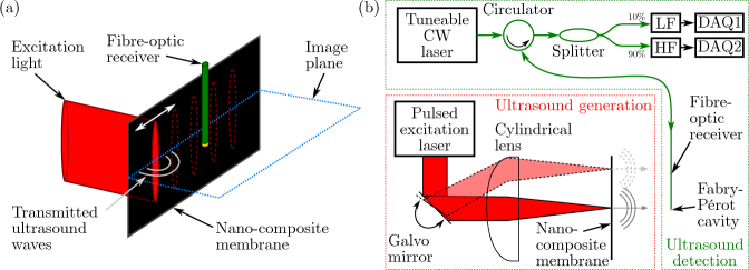
Fig. 1.
Optical ultrasound generation and detection. (a) Using a cylindrical lens, excitation light was delivered to a small, eccentric area of an optically absorbing membrane comprising a nano-composite. Ultrasound was generated photoacoustically in this area, and a linear acoustic aperture was scanned sequentially by translating the focal spot across the membrane using a galvo mirror. (b) Schematic of the set-up used to optically generate (red box) and detect (green box) ultrasound. A fibre-optic acoustic receiver, comprising a Fabry-Pérot cavity fabricated at its tip, was interrogated using a tuneable continuous wave laser. Using an optical splitter, 10% of the reflected light was recorded using a low frequency photodiode to record the cavity transfer function in order to identify the resonance wavelength; the remaining 90% was coupled into a high-frequency photodiode to record the acoustic signal. CW: continuous-wave; LF/HF: low-/high-frequency photodiode; DAQ: data acquisition.

