Fig. 3.
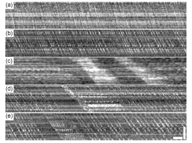
Characteristic spatiotemporal traces of the erythrocytes in retinal capillaries obtained from the AONCO imaging. (a) Type 1, the blood flow in the capillary contained densely packed erythrocytes flowing in non-single files and no leukocytes. (b) Type 2, the blood flow contained erythrocytes flowing in single files and no leukocytes. (c) Type 3a, the blood flow contained both leukocytes (hypo-reflective bands) and non-single file erythrocyte aggregation (hyper-reflective bands). (d) Type 3b, the blood flow contained both leukocytes (hypo-reflective bands) and light erythrocyte aggregation (hyper-reflective bands). (e) Type 4, the blood flow contained leukocytes accompanied by non-aggregated upstream erythrocytes. All spatiotemporal plots were extracted from retinal images acquired at 800 fps, with the imaging light focused on the capillary bed. The temporal (horizontal) scale bar indicates 25 ms and the spatial (vertical) scale is 25 µm.

