Figure 2.
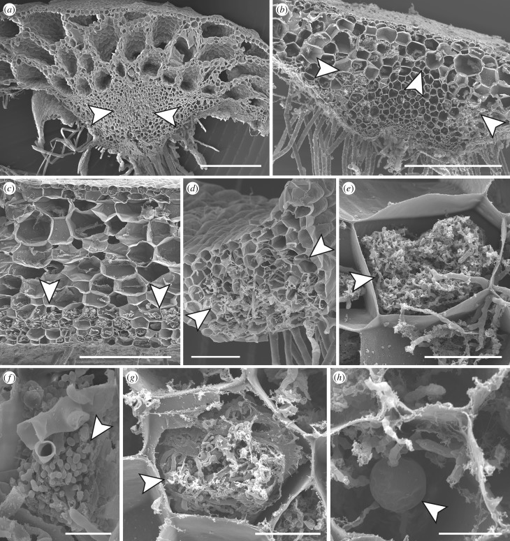
Cytology of Glomeromycotina associations in liverworts. Scanning electron micrographs of cross sections through thalli of (a) Neohodgsonia mirabilis; (b,f,g) Dumortiera hirsuta; (c,e) Monoclea forsteri; (d) Fossombronia foveolata; (f) Marchantia pappeana. Fungal colonization usually occupies the thallus central midrib (arrowed) (a,d). In some Marchantiopsida liverworts colonization is sometimes confined to a region overarching the midrib hyaline strand (arrowed) (b), or is restricted to the thallus ventral cell layers (arrowed) (c). Fungal structures include arbusculated coils (arrowed) (e), arbuscules terminal on trunk hyphae (arrowed) (f), coils (arrowed) (g), and vesicles of varying diameters (arrowed) (h). Scale bars: (a–c) 500 µm; (d) 100 µm; (e) 50 µm; (g,h) 20 µm; (f) 10 µm.

