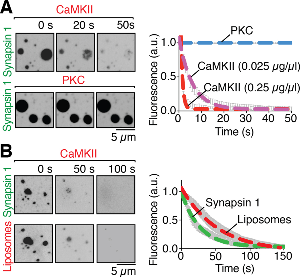
Fig. 4. Phosphorylation of the intrinsically disordered region of synapsin 1 disperses condensates of either synapsin 1 alone or synapsin 1 and liposomes.
(A) Left: fluorescence images of EGFP-Synapsin 1 condensates preincubated with either CaMKII (0.025 μg/μl), calmodulin and calcium, or PKC, PS, DAG and calcium, upon addition, at 0 seconds, of ATP (200 μM), demonstrating dispersion of synapsin by CaMKII, but not by PKC. Right: time course of the effect of the kinases on the condensates, as assessed by the decrease of florescence on ROIs corresponding to randomly selected droplets (B) Left: Fluorescence images of liposome-synapsin condensates preincubated with CaMKII (0.25 μg/μl), calmodulin and calcium, upon addition, at 0 seconds, of ATP (200 μM), demonstrating dispersion of both synapsin and liposomes. Right: Time-course of the effect of CaMKII on liposome-synapsin droplets dispersion. Error bars represent s.e.m.; dashed lines represent the fit with a single exponential function. Incubations were carried out at RT in a buffer of physiological salt concentration supplemented with 3% PEG 8,000.

